Welcome
Contents
Events
Article sur le centre MEG dans la Marseillaise, dans le cadre de l'inauguration de l'ILCB File:Article ILCB la Marseillaise.pdf
Formation Connectivité avec Fieldtrip 21-22 novembre 2016 File:Fiche annonce - Connectivité en MEG et EEG - 2016.pdf http://www.fieldtriptoolbox.org/workshop/marseille2016b
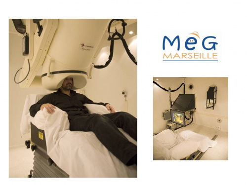
The Marseille MEG platform.
Magnetoencephalography (MEG) consists in recording brain magnetic activity. This activity is the counterparts of the electrical activity that originate from the brain, the well-known EEG. These techniques are the only ones that are directly related to the neural activity and have enough time resolution to track brain activity. One of the great advantages of the MEG other EEG is the very small effect of the geometry of the different media of the head, especially the skull, on the signal recorded compared to EEG.
The MEG laboratory of Marseille uses a 248 magnetometers MEG system (4D Neuroimaging magnes 3600), installed within the Neurophysiology department of Timone hospital (Head: F. Bartolomei). The system was co-financed by: Conseil Régional PACA, Conseil Général 13, Conseil Général 06, Marseille Provence Métropole, INSERM, CNRS, INRIA. It is currently co-financed by Aix-Marseille Université (though France Life Imaging) and INSERM.
The laboratory is equipped with stimulation apparatus that includes video projection and Stax calibrated system for auditory stimulation. It belongs to the Aix-Marseille University and is accessible to all teams involved in fundamental and clinical brain research.
The research team of the MEG laboratory is specialized in confronting results of source localization to intracerebral EEG (Dubarry et al 2014, Gavaret et al 2004, Koessler et al 2010), and in designing and optimizing signal processing methods for multimodal functional investigation of human cerebral activity (pathological and physiological) (Jmail et al 2011, Krieg et al 2011).
The AnyWave Software
The MEG laboratory is currently developping a software called Anywave for the visualisation and analysis of electrophysiological data: MEG, EEG, SEEG (developper: Bruno Colombet, with help from M. Woodman, Engineer financed by Fondation pour la recherche medicale).
The guiding principles are modularity and cross-platform portability.
See AnyWave.
Staff
Jean-Michel Badier, technical director
Christian Bénar, scientific director
Sophie Chen, engineer
Bruno Colombet, software developer
Publications
Cognition
Spatio-temporal dynamics of morphological processing in visual word recognition. Cavalli E, Colé P, Badier JM, Zielinski C, Chanoine V, & Ziegler JC. J Cogn Neurosci. 2016 Aug;28(8):1228-42.
On the Cortical Dynamics of Word Production : A Review of the MEG Evidence. Munding, D., Dubarry, A.-S., & Alario, F.-X. Lang Cogn Neurosci. 2016.
MEG studies of word production: What next? Munding, D., Dubarry, A.-S., & Alario, F.-X. Lang Cogn Neurosci. 2016.
Characterization of Cortical Networks and Corticocortical Functional Connectivity Mediating Arbitrary Visuomotor Mapping. Brovelli A, Chicharro D, Badier JM, Wang H, Jirsa V. J Neurosci. 2015 Sep 16;35(37):12643-58.
Clinical research
Comparison of Brain Networks During Interictal Oscillations and Spikes on Magnetoencephalography and Intracerebral EEG. Jmail N, Gavaret M, Bartolomei F, Chauvel P, Badier JM, Bénar CG. Brain Topogr. 2016 Sep;29(5):752-65.
Ictal Magnetic Source Imaging in Presurgical Assessment. Badier JM, Bénar CG, Woodman M, Cruto C, Chauvel P, Bartolomei F, Gavaret M. Brain Topogr. 2016 Jan;29(1):182-92.
Magnetic source imaging in posterior cortex epilepsies. Badier JM, Bartolomei F, Chauvel P, Bénar CG, Gavaret M. Brain Topogr. 2015 Jan;28(1):162-71.
Interictal networks in magnetoencephalography. Malinowska U, Badier JM, Gavaret M, Bartolomei F, Chauvel P, Bénar CG. Hum Brain Mapp. 2014 Jun;35(6):2789-805.
MEG and EEG sensitivity in a case of medial occipital epilepsy. Gavaret M, Badier JM, Bartolomei F, Bénar CG, Chauvel P. Brain Topogr. 2014 Jan;27(1):192-6
methodological developments
AnyWave: A cross-platform and modular software for visualizing and processing electrophysiological signals. Colombet B, Woodman M, Badier JM, Bénar CG. J Neurosci Methods. 2015 Jan 19;242C:118-126.
Organic electrochemical transistors for clinical applications. Leleux P, Rivnay J, Lonjaret T, Badier JM, Bénar C, Hervé T, Chauvel P, Malliaras GG. Adv Healthc Mater. 2015 Jan;4(1):142-7.
Simultaneous recording of MEG, EEG and intracerebral EEG during visual stimulation: from feasibility to single-trial analysis. Dubarry AS, Badier JM, Trébuchon-Da Fonseca A, Gavaret M, Carron R, Bartolomei F, Liégeois-Chauvel C, Régis J, Chauvel P, Alario FX, Bénar CG. Neuroimage. 2014 Oct 1;99:548-58.
Conducting polymer electrodes for electroencephalography. Leleux P, Badier JM, Rivnay J, Bénar C, Hervé T, Chauvel P, Malliaras GG. Adv Healthc Mater. 2014 Apr;3(4):490-3.
